We present examples of how hyperspectral cameras are used in the beauty and medical fields, including spectral measurements at the cellular level via microscope connections and the visualization of various physiological data.
Microscope-Connected Photography (Pathology Specimens)
FRET Bioimaging (Macroscopic Observation of Fluorescence Resonance Energy Transfer)
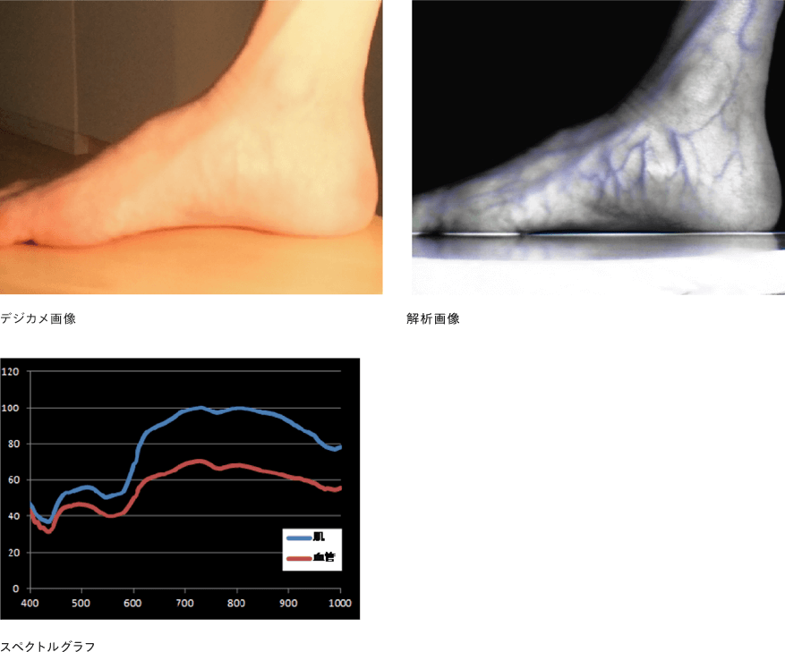
Vessel Detection
Multidimensional Analysis of Biometric Data
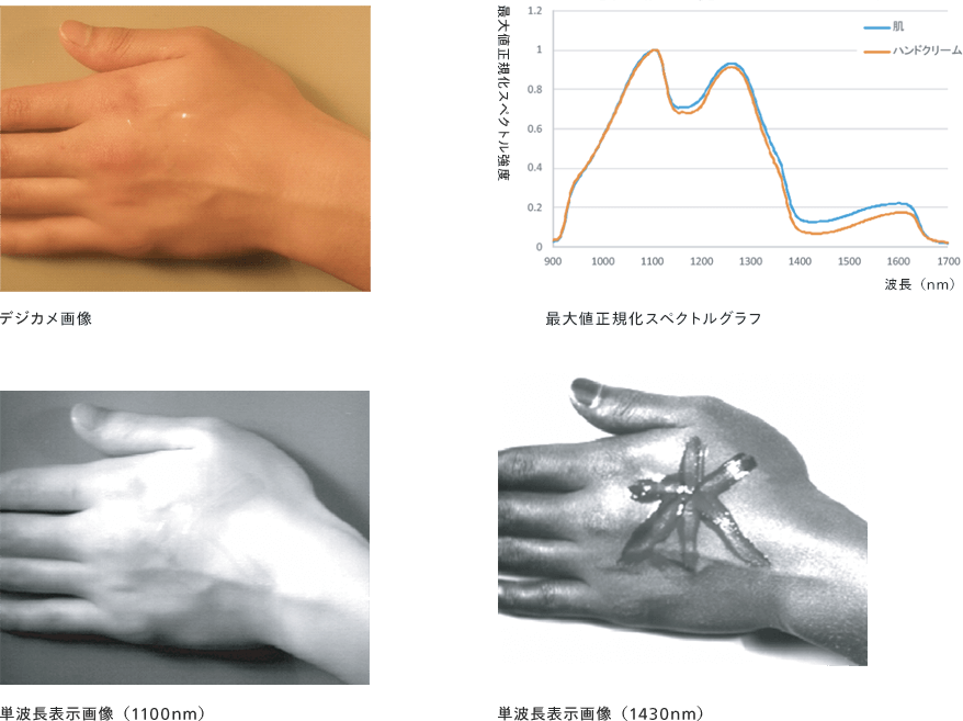
Photo shoot for a moisturizing cream
Microscope-Connected Photography (Pathology Specimens)

By connecting a hyperspectral camera to a microscope, spectral measurements can be performed at the cellular level, as described above.
FRET Bioimaging (Macroscopic Observation of Fluorescence Resonance Energy Transfer)
Quantitative imaging of PKA activity
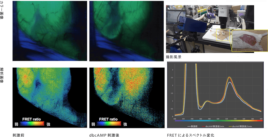
We successfully performed macroscopic measurements of PKA activation in the thigh muscles of genetically modified mice that visualize Protein Kinase A (PKA) activation following dCAMP stimulation.
Vessel Detection
Multidimensional Analysis of Biometric Data

Biological spectral imaging performs spectral analysis for each spatial pixel, enabling the visualization of various biological features (blood vessels, wrinkles, and skin texture). The application of this technology makes multidimensional analysis of biological information possible.
Photo shoot for moisturizing cream
APPLICATION
Application Fields
EBA Spectral Technology is used in a wide range of fields.


CONTACT
Contact Us
For telephone inquiries
03-6433-1517[Business Hours] Weekdays 10:00-18:00